Orthognathic treatment after removal of extensive mandibular osteoma
Orthognathic treatment after removal of extensive osteosarcoma of the mandible
Osteoma, or osteoma, is a benign, slow-growing cancerous lesion that develops in bone tissue. Osteomas are located primarily in the pelvis, shoulder blades, in the epiphyses of long bones (femoral and tibial), as well as in the phalanges. The location of the osteoma is also often the craniofacial bones.
Ossificosis is often, but not always, characterized by the absence of painful symptoms and proliferation of spongy substance (in the case of the face – most often in the paranasal sinuses, in the maxilla and mandible). The developing osteotoma over time takes the place of normal, mature bone tissue – it replaces it with tissue of much greater hardness (numerous bone beams and a small amount of bone marrow), deforming the shape of the involved bone. In the absence of pain and small size (e.g. up to 1.5 cm) the osteoma in many cases is not diagnosed at all.
There are several methods of treating osteomas. They are usually asymptomatic and no action is taken in such a case (only routine X-ray follow-up is indicated). In other cases, non-steroidal anti-inflammatory drugs (NSAIDs), spooning, percutaneous radiofrequency current ablation or surgical treatment may be used.
The occurrence of multiple ossicles is a characteristic symptom of one of the variants of the syndrome of familial adenomatous polyposis of the large intestine – Gardner syndrome.
The paper by the authors from the Department of Oral and Maxillofacial Surgery at Istanbul University presents the case of a 17-year-old patient with an extensive osteoma that developed in the condylar process of the mandible on the left side. The lesion, which had been developing for the previous 4 years, caused the patient’s facial asymmetry. However, the osteoma did not give pain; among the symptoms, the patient reported mainly problems with swallowing (dysphagia).
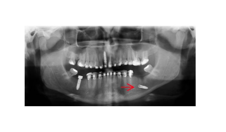
Extraoral examination reveals a subcutaneous, hard and convex mass on the left side of the mandible and a shift of the mandible to the right, noticeable when opening the mouth. Intraoral examination showed no signs of pathology in the mandibular branch. The pantomographic image taken showed an extensive infiltrative mass on the left side of the mandible, impermeable to X-rays. These findings suggest a calcified odontogenic tumor requiring segmental or block resection. A CBCT scan was performed to further evaluate the nature and extent of the osteotoma – this scan showed a diffuse lesion measuring 4.5 × 3.5 × 3 cm in the left shoulder of the mandible. Due to the presence of dysphagia, facial asymmetry and progression of the lesion, the decision was made to perform surgical correction in the mandible on the left side.
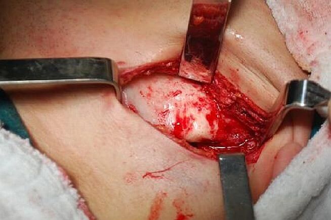
The patient was administered general anesthesia (articaine hydrochloride epinephrine – 6 ml 1 / 100,000) with nasotracheal intubation and cut from an extraoral access. Using a slit drill and chisel, a flap was cut in the lateral and medial osteoma, while giving the mandible a normal shape. The healing process proceeded without complications. A post-operative follow-up 6 months later using dental tomography showed no pathology. There has been no recurrence of the osteoma in the following 5 years.
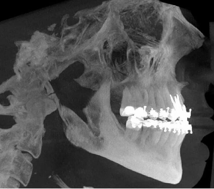
The patient – in addition to osteoma – also had a Class III bone defect, diagnosed by cephalometric analysis. Therefore, the decision was made to start orthodontic treatment. After initial treatment, the patient had third molars removed, then the patient was prepared for orthognathic surgery. It was performed 7 years after the removal of the osteotomy – when the operated mandible was fully healthy and the man had reached an age appropriate for this type of surgery. LeFort I osteotomy in the maxilla was performed 3 mm anteriorly and 4 mm to the right side. On the other hand, an orthognathic procedure for bilateral sagittal osteotomy of the mandible was performed, achieving a displacement of 1 mm proximally and 2 mm to the left. Less than a year after surgery, the patient’s missing teeth were replaced with dental implants.
Article title: Orthognathic Surgery after Mandibular Large-Volume Osteoma Treatment
Sabit Demircan1, Sabri Cemilişler 2, Aydın Gümüşdal 2, Begüm Genç 2
1 Private Practice, Beykent University Vocational School, Turkey
2 Istanbul University Faculty of Dentistry Oral and Maxillofacial Surgery Department, Turkey